
Author:admin
Time:2020-10-29 13:16:23
Views:
Multiple sclerosis (MS) is an autoimmune disease characterized by white matter inflammatory demyelinating lesions of the central nervous system. The most common sites involved in the disease are periventricular white matter, optic nerve, spinal cord, brainstem and cerebellum. The main clinical features are multiple lesions scattered in the white matter of the central nervous system and remission and recurrence in the course of the disease. multiple symptoms and signs in space and time in the course of the disease. The main function of oligodendrocytes is to wrap axons in the central nervous system, form an insulated myelin sheath structure, assist the jumping transmission of bioelectric signals, and maintain and protect the normal function of neurons.
The dysfunction of oligodendrocytes (OLs) is regarded as one of the major causes of inefficient remyelination in multiple sclerosis, resulting gradually in disease progression. Oligodendrocytes are derived from oligodendrocyte progenitor cells (OPCs), which populate the adult central nervous system, but their physiological capability to myelin synthesis is limited. The low intake of essential lipids for sphingomyelin synthesis in the human diet may account for increased demyelination and the reduced efficiency of the remyelination process。
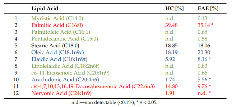
In our study on lipid profiling in an experimental autoimmune encephalomyelitis brain, we revealed that during acute inflammation, nervonic acid synthesis is silenced, which is the effect of shifting the lipid metabolism pathway of common substrates into proinflammatory arachidonic acid production. In the experiments on the human model of maturating oligodendrocyte precursor cells (hOPCs) in vitro, we demonstrated that fish oil mixture (FOM) affected the function of hOPCs, resulting in the improved synthesis of myelin basic protein, myelin oligodendrocyte glycoprotein, and proteolipid protein, as well as sphingomyelin
The lipid composition of experimental autoimmune encephalomyelitis EAE and healthy control (HC) brains. Red color indicates the lipids that are reduced in the course of disease. A blue color indicates the lipids that are relatively upregulated during EAE, while a green color indicates the new appearing lipids involved in arachidonic acid (AA) metabolism. Data are presented as the percentage of lipids from three independent experiments (one mouse per one experiment)

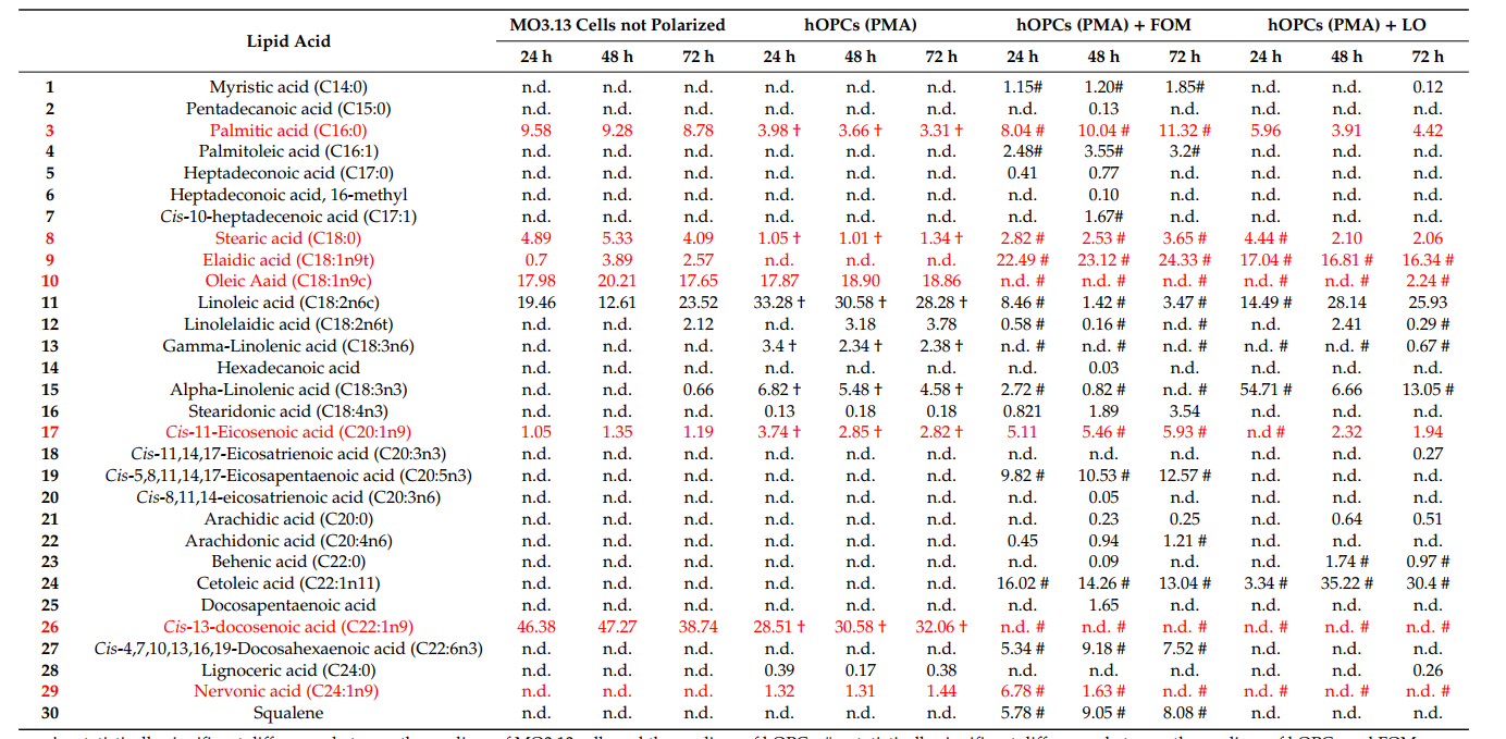
Next, we examined whether supplementation of the culture medium with FOM, in comparison to LO, would affect the lipid acid esters that are bound to sphingosine, creating sphingomyelin in the maturing hOPCs. PMA-stimulated MO3.13 cells (hOPCs) were incubated alone or in the presence/absence of FOM at a final concentration of 5% or LO used as a control for 72 h. In the group of detected lipids, we noted three of the most important lipid acid esters being substrates for the synthesis of sphingomyelin: nervonic, palmitic, and stearic acid (Table 2). We found at 24 h of incubation that the percentage of NA ester in hOPCs was increased six to seven times in the presence of 5% FOM, in comparison to hOPCs incubated alone. The effect of FOM and linseed oil (LO) on the lipid composition of maturing hOPC. The red color indicates the main lipids that are involved in NA synthesis or directly bind to sphingomyelin during hOPC maturation. Data are presented as the percentage of lipids from four independent experiments
Therefore, using a cytokine multiple profiling assay, we have analyzed supernatant concentrations of 27 cytokines, chemokines, and growth factors. We revealed that hOPCs cultured with FOM in comparison to hOPCs cultured in medium produced statistically significant increased amounts of fibroblast growth factor 2 (FGF-2) and vascular endothelial growth factor (VEGF), as well as decreased amounts of interleukin (IL)-5, IL-6, IL-7, IL-8, IL-15, IL-17, eotaxin, granulocyte colony-stimulating factor (G-CSF), interferon (IFN)-γ, monocyte chemoattractant protein 1 (MCP-1). Additionally, FOM reduces proinflammatory cytokines and chemokines, and enhances fibroblast growth factor 2 (FGF2) and vascular endothelial growth factor (VEGF) synthesis by hOPCs was also demonstrated.

In this study, we demonstrated that remyelination can be affected by the presence of fish oils rich in EPA, DHA, and NA in the microenvironment. We found that maturing hOPCs cultured in the presence of FOM incorporated and metabolized NA, and transformed into OLs characterized by normal morphology and prolonged lifespan. Based on these observations, we propose that the intake of FOM rich in the nervonic acid ester may improve OL function, affecting OPC maturation and limiting inflammation
The content of NA is high in the brain and nerve tissue, and it can act on all aspects of brain development, including promoting the proliferation and differentiation of brain nerve cells. through the synthesis of glycosphingolipids (cerebral glycosides, gangliosides, etc.) and sphingomyelin in the human body, to regulate and improve the composition, structure and function of the biofilm. It can regulate the level of neurotransmitters in the brain, make the information transmission between nerve cells faster and more accurate, promote the recognition of cells, including cell regeneration and contact inhibition, and can restore the activity of nerve endings, improve brain function, and enhance people's attention and memory.